The retina works a little like a camera sensor built from two different pixel types: rods that pick up faint light and cones that handle colour and daylight detail. Retinitis Pigmentosa is what happens when the rod pixels begin failing first, in a pattern that is broadly predictable across patients even though the speed of change is not. It remains the leading inherited driver of gradual vision loss worldwide, and that is precisely why the term keeps resurfacing once an ophthalmologist in Uganda first mentions it to a family.
No two RP patients travel the same road at the same pace, so before any treatment discussion begins, we need a clear read on exactly how much ground has already been covered. That single data point — the current stage of disease — tends to steer nearly every recommendation that comes afterward.
10+
years Experience
years Experience
5000+
Patients treated
Patients treated
100+
Countries served
Countries served
NABH
Guided facility
Guided facility
Ocular Symptoms
Because rods fail first, difficulty seeing after sunset or inside a poorly lit room is usually the earliest complaint, sometimes noticed years before anything else changes. The visual field then begins narrowing from the outside in, producing the tunnel-like vision RP is commonly associated with, occasionally with a ring-shaped gap appearing partway through that process. Central, cone-driven vision tends to hold out the longest, but ‘longest’ is not the same as ‘forever’ — it can eventually be affected too, once the disease has advanced further.
Underlying Causes
TRP is not caused by one faulty gene but by well over 80 of them, with RHO, USH2A, and RPGR turning up most frequently. The specific gene involved decides how the condition moves through a bloodline — dominant in some families, recessive in others, and X-linked in a third pattern altogether. Where a Ugandan household has multiple affected members, mapping that inheritance pattern across the family often reveals nearly as much as the eye examination itself.
Diagnosis for Ugandan Patients
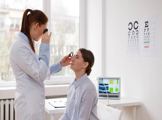
Four investigations carry most of the diagnostic weight: full-field electroretinography, which records the electrical signal generated by rods and cones; OCT scanning, which images the surviving photoreceptor layers; Goldmann visual field testing, which maps precisely how far peripheral vision has retreated; and fundus autofluorescence, which outlines the border between healthy and degenerating retina. Genetic testing is offered whenever feasible, since the particular gene involved can reshape both the outlook and the treatment options worth pursuing.
Meet The Team
Our Eye Specialists

Dr. Pawan
Associate Professor, Vitreo-Retina Surgeon. 10+ Years Experience
Retinal disorders, diabetic retinopathy, and advanced retina procedures. Provides diagnostic evaluation and treatment planning for international patients.

Dr.Pallavi
Senior Consultant Ophthalmologist & Eye Surgeon· 26+ yrs experience
Areas of Focus: Cataract surgery, corneal disorders, refractive procedures, and glaucoma management. Provides comprehensive eye care and surgical expertise for global patients.
Dr. Robin
Consultant Ophthalmologist & Eye Surgeon, MD, DO .16+ Years Experience
Areas of Focus: Cataract surgery, corneal disorders, refractive procedures, and glaucoma management. Provides comprehensive eye care and surgical expertise for global patients.
Treatment Approach in India
Where a patient’s assessment supports it, regenerative stem cell therapy forms one piece of a broader plan built around preserving remaining retinal function. That plan typically runs alongside low-vision rehabilitation, guidance on safer lighting and mobility, and nutritional advice — so a patient travelling from Uganda leaves with an ongoing management strategy rather than a single isolated procedure.
Reviews
What Our Patients Say

DR NAAGALAKSHMI R
2 months ago

Asha Babbar
2 months ago
The overall experience at the RP Treatment Center was very good. The staff was supportive and caring, and we provided proper attention and guidance during treatment. The environment at the center was clean and comfortable. The doctors and staff tried to understand patients' needs and explained the treatment process well.
Overall, I am satisfied with the service and care. Highly recommended!
Overall, I am satisfied with the service and care. Highly recommended!

Harshita Kapoor
2 months ago
Life‑Changing Experience with RP Therapy”
After years of struggling with vision loss due to Retinitis Pigmentosa, I finally found hope here. The doctors were highly professional, explained the stem cell treatment in detail, and gave me confidence at every step. The therapy was smooth, and I’ve already noticed positive changes in my daily life. Their dedication, care, and advanced medical approach make this center truly trustworthy
After years of struggling with vision loss due to Retinitis Pigmentosa, I finally found hope here. The doctors were highly professional, explained the stem cell treatment in detail, and gave me confidence at every step. The therapy was smooth, and I’ve already noticed positive changes in my daily life. Their dedication, care, and advanced medical approach make this center truly trustworthy

Yash Chauhan
2 months ago
I had a really good experience at RP Treatment Centre. The doctor listened to my problem patiently and explained the treatment in a simple way. The staff was polite and the clinic was clean. I started feeling better after the treatment. Highly recommended!

pragya chauhan
2 months ago
Very satisfied with the service. The doctor is experienced and gives proper attention to every patient. The staff is friendly, and the overall environment is comfortable. Thank you, RP Treatment Centre, for the excellent care.

Manisha kapoor
2 months ago
I had been struggling with Retinitis Pigmentosa for years, and finding this center was truly life‑changing. The doctors provided a thorough medical evaluation and explained every step of the stem cell therapy process with clarity. The treatment was smooth, professional, and I noticed encouraging improvements in my vision within weeks. Their continuous follow‑up care and compassionate support made me feel confident throughout the journey. Highly recommended for anyone seeking advanced RP treatment in India.
Frequently Asked Questions
Will RP eventually leave me completely blind?
“Not necessarily — outcomes differ substantially from person to person, largely depending on which gene is responsible and how it is inherited. Many people with RP keep meaningful, usable vision, especially centrally, well into later life. That variability is exactly why every case is assessed individually rather than assumed to follow one fixed path.”
Two of my children already show early signs — should we be concerned about our other children?
”
That depends on the inheritance pattern tied to the specific gene at fault, and genetic testing is the most dependable way to work that out. We can talk you through what that likely means for your family, though for decisions around future children we would recommend involving a certified genetic counsellor as well.”
Can’t a local eye clinic in Uganda simply test for RP?
“A routine eye check is built to catch things like refractive error or cataract, not RP. Confirming and staging RP calls for equipment — ERG and detailed field mapping in particular — that most general clinics don’t stock, which is why a focused evaluation usually means travelling somewhere it is available.”
What should we bring to the first consultation?
“Bring along any existing eye reports, a general sense of your family’s eye-health history, and a rough timeline of when symptoms began and how they have shifted since. Even that basic background can speed up the diagnostic process considerably.”