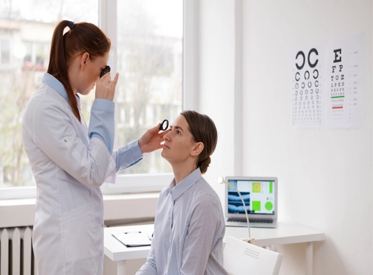
Picture three retinal structures — the choroid, the retinal pigment epithelium sitting just above it, and the photoreceptors resting on top of that — wasting away in lockstep rather than one at a time. That’s choroideremia, and because the responsible gene sits on the X chromosome, the condition turns up almost exclusively among boys and men; women carrying the gene usually get by with only subtle changes that rarely interfere with everyday sight.
For families arriving from Rwanda, the path to this diagnosis often begins with a routine fundus photograph on a male relative that a local eye doctor recognised as different from the usual RP pattern — different enough to warrant a genetic test confirming choroideremia specifically, rather than settling for a broader retinal-dystrophy label.
10+
years Experience
years Experience
5000+
Patients treated
Patients treated
100+
Countries served
Countries served
NABH
Guided facility
Guided facility
Ocular Symptoms
Trouble seeing after dark, starting in childhood, is nearly always the opening sign, showing up well before anything else does. The visual field then contracts steadily over the teenage years and beyond. What genuinely marks choroideremia out is the sheer length of time central vision keeps functioning normally — frequently decades longer than in comparable inherited retinal conditions — although it is not indefinite, and can be lost once the macula itself becomes involved, usually somewhere in mid-to-late adulthood.
Underlying Causes
A faulty CHM gene, carried on the X chromosome, is behind the condition, leaving retinal cells short of the REP1 protein they depend on to stay alive and functioning. Since it’s passed down through the X chromosome, a carrier mother has roughly even odds of passing the mutation to each son, who will be affected, and roughly even odds of passing carrier status to each daughter.
Diagnosis for Rwandan Patients
A scalloped, moth-eaten look to the retina on fundus examination is a fairly telling sign of choroideremia, and paired with OCT scans and electroretinography, it’s usually enough to point away from other rod-cone dystrophies fairly quickly. A CHM genetic test settles the matter conclusively and is especially useful for checking whether female relatives are carriers.
Meet The Team
Our Eye Specialists

Dr. Pawan
Associate Professor, Vitreo-Retina Surgeon. 10+ Years Experience
Retinal disorders, diabetic retinopathy, and advanced retina procedures. Provides diagnostic evaluation and treatment planning for international patients.

Dr.Pallavee
Senior Consultant Ophthalmologist & Eye Surgeon· 26+ yrs experience
Areas of Focus: Cataract surgery, corneal disorders, refractive procedures, and glaucoma management. Provides comprehensive eye care and surgical expertise for global patients.
Dr. Robin
Consultant Ophthalmologist & Eye Surgeon, MD, DO .16+ Years Experience
Areas of Focus: Cataract surgery, corneal disorders, refractive procedures, and glaucoma management. Provides comprehensive eye care and surgical expertise for global patients.
Treatment Approach in India
With central vision holding on for so long in most cases, treatment is built around protecting that window for as long as possible — weighing regenerative stem cell therapy for suitable candidates, alongside regular monitoring of disease progression and early planning for low-vision support ahead of any eventual changes centrally.
Reviews
What Our Patients Say

DR NAAGALAKSHMI R
2 months ago

Asha Babbar
2 months ago
The overall experience at the RP Treatment Center was very good. The staff was supportive and caring, and we provided proper attention and guidance during treatment. The environment at the center was clean and comfortable. The doctors and staff tried to understand patients' needs and explained the treatment process well.
Overall, I am satisfied with the service and care. Highly recommended!
Overall, I am satisfied with the service and care. Highly recommended!

Harshita Kapoor
2 months ago
Life‑Changing Experience with RP Therapy”
After years of struggling with vision loss due to Retinitis Pigmentosa, I finally found hope here. The doctors were highly professional, explained the stem cell treatment in detail, and gave me confidence at every step. The therapy was smooth, and I’ve already noticed positive changes in my daily life. Their dedication, care, and advanced medical approach make this center truly trustworthy
After years of struggling with vision loss due to Retinitis Pigmentosa, I finally found hope here. The doctors were highly professional, explained the stem cell treatment in detail, and gave me confidence at every step. The therapy was smooth, and I’ve already noticed positive changes in my daily life. Their dedication, care, and advanced medical approach make this center truly trustworthy

Yash Chauhan
2 months ago
I had a really good experience at RP Treatment Centre. The doctor listened to my problem patiently and explained the treatment in a simple way. The staff was polite and the clinic was clean. I started feeling better after the treatment. Highly recommended!

pragya chauhan
2 months ago
Very satisfied with the service. The doctor is experienced and gives proper attention to every patient. The staff is friendly, and the overall environment is comfortable. Thank you, RP Treatment Centre, for the excellent care.

Manisha kapoor
2 months ago
I had been struggling with Retinitis Pigmentosa for years, and finding this center was truly life‑changing. The doctors provided a thorough medical evaluation and explained every step of the stem cell therapy process with clarity. The treatment was smooth, professional, and I noticed encouraging improvements in my vision within weeks. Their continuous follow‑up care and compassionate support made me feel confident throughout the journey. Highly recommended for anyone seeking advanced RP treatment in India.
Frequently Asked Questions
Why does choroideremia mainly show up in men?
It’s simply how X-linked inheritance works. Men have a single X chromosome, so one faulty CHM copy is all it takes to cause the disease. Women have two, and the working copy carriers still have is generally enough to spare them meaningful vision loss.
As a carrier myself, is it certain my sons will be affected?
No — the odds work out to roughly one in two for each son to inherit the mutation, and roughly one in two for each daughter to become a carrier. A genetic test can put a firmer number on that for your own family.
Would an eye doctor be able to spot this instead of standard RP just by looking?
Frequently, yes. The scalloped thinning across both the choroid and RPE gives choroideremia a somewhat different look on examination than the more even photoreceptor loss typical of RP, though a genetic test is still what confirms it beyond doubt.
How soon should treatment or monitoring conversations start?
The sooner the better, generally, since it leaves the most options open while central vision remains sharp. We’d much rather assess a patient well before central-vision symptoms appear than after.