
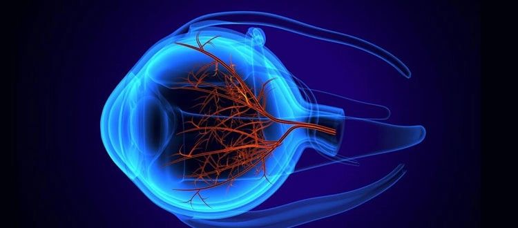
02 Nov How to Get Tested for Retinitis Pigmentosa: Step-by-Step from First Symptoms to Diagnosis
Last updated: 2 November 2026 | Author: Prashant Tyagi (Stem Cell Biotechnology Specialist, 10+ years) and Dr. Pallavee Senior Consultant | Ophthalmologist & Eye Surgeon 22+ Years Experience
Trouble seeing in dim light, bumping into things at night or noticing that your side vision seems to be narrowing can be early signs of retinitis pigmentosa (RP). Many people wait months or years before getting checked, partly because the symptoms come on slowly. This guide explains the path from first symptoms to a confirmed diagnosis: who to see, which tests are done, what genetic testing can and cannot tell you, and what to do once you have an answer.
Quick Navigation
- Early symptoms to notice
- Step 1: See an eye specialist
- Step 2: Core eye tests
- Step 3: Specialised tests
- Step 4: Genetic testing
- Conditions that can look similar
- After diagnosis
- Testing family members
- Frequently asked questions
- Checklist
Early Symptoms to Notice
- Difficulty seeing in dim light or at night (night blindness), often the first sign
- Slow adjustment when moving from bright to dark places
- Loss of side vision, so that vision feels like looking through a tube
- Tripping over objects or struggling in crowds
- Sensitivity to glare
- In later stages, reduced central vision and colour changes
RP usually affects both eyes and progresses slowly, at different speeds in different people. Our overview page on retinitis pigmentosa explains the condition in more detail. Hearing problems alongside RP can point to Usher syndrome, so mention any hearing difficulty to your doctor.
Step 1: See an Eye Specialist
Start with an ophthalmologist, ideally a retina specialist. Bring your history: when symptoms began, how they have changed, family history of eye disease or early vision loss, any hearing problems and the medicines you take. Dilated examination of the back of the eye allows the doctor to see the typical retinal changes, such as pigment clumps and thinned blood vessels, though they may be subtle early on.
Step 2: Core Eye Tests
| Test | What it shows in RP |
|---|---|
| Visual acuity and refraction | Sharpness of vision and whether glasses help |
| Dilated fundus examination and photographs | Retinal changes and a record for comparison |
| Visual field test | The ring-shaped or narrowing field typical of RP |
| OCT (optical coherence tomography) | Retinal layer thinning and swelling at the macula |
| Fundus autofluorescence | Rings or patterns showing where retinal cells are under stress |
Step 3: Specialised Tests
The electroretinogram (ERG) is the key confirmatory test. It measures the electrical response of the rods and cones to light. In RP, the rod response is usually reduced or absent early, even when the retina still looks nearly normal. Dark adaptation testing may also be done. These tests are available at larger eye hospitals, so your ophthalmologist may refer you.
Step 4: Genetic Testing
RP is usually inherited, and over a hundred genes have been linked to retinal dystrophies. Genetic testing can:
- Confirm the diagnosis and separate RP from similar conditions
- Show the inheritance pattern (dominant, recessive or X-linked), which helps family planning
- Identify the small group of patients who may qualify for an approved gene therapy or a clinical trial
Genetic testing does not always find the cause, and a negative result does not rule out RP. Ask for genetic counselling before and after testing so you understand what the result can and cannot mean. You can look up registered trials on the Clinical Trials Registry – India.
Conditions That Can Look Similar
A careful workup separates RP from other conditions, including cone dystrophy, rod-cone dystrophy, Leber congenital amaurosis, choroideremia, Bardet-Biedl syndrome, vitamin A deficiency and the effects of some medicines. The distinction matters, as the outlook and the options differ.
After Diagnosis
- Regular follow-up: repeat visual fields and OCT, usually yearly, to track the pace of change.
- Treat complications: cataract and macular swelling are common in RP and can often be treated, improving vision.
- Protect the eyes: UV-filtering sunglasses and avoiding smoking are widely advised.
- Low vision support: mobility training, lighting and assistive technology help you stay independent.
- Emotional support: a diagnosis is a lot to take in. Talk to family, a counsellor or a patient group.
No treatment can currently cure RP. An approved gene therapy exists only for a small group with a specific gene change. Stem cell approaches for RP are investigational, and results vary. They should never replace regular care, and anyone promising a cure should be questioned closely. See our page on retinitis pigmentosa treatment in India for how options are discussed.
Testing Family Members
Because RP is often inherited, relatives may wish to be checked, particularly children of an affected parent. A baseline eye examination, with OCT and a visual field where age permits, is a reasonable start. Genetic counselling can explain each person’s risk.
Frequently Asked Questions
At what age is RP usually diagnosed?
Often in adolescence or early adulthood, but it can appear earlier or later depending on the type.
Can a routine eye test detect RP?
A standard sight test alone may miss early RP. A dilated retinal examination, visual field and ERG give a more reliable answer.
Is genetic testing necessary?
It is not required to diagnose RP, but it is strongly recommended because it guides counselling and any trial options.
Will RP always lead to blindness?
Not always. Many people keep useful vision for decades. The course varies widely between individuals.
Can I send my reports for review?
Yes. Use the contact page to share your reports, or read more on our blog.
Checklist
- Noted symptoms, with dates, and family history
- Seen an ophthalmologist, ideally a retina specialist
- Had a dilated examination, OCT and visual field
- Asked about ERG and genetic testing
- Considered genetic counselling
- Checked hearing if relevant
- Planned yearly follow-up
- Learned that no cure exists and that claims should be questioned
More Official Sources
- National Eye Institute (NEI)
- American Academy of Ophthalmology (AAO)
- MedlinePlus Genetics
- Foundation Fighting Blindness
- Clinical Trials Registry – India (CTRI)
- A Closer Look at Stem Cells (ISSCR patient resource)

Sorry, the comment form is closed at this time.