
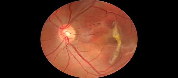
11 Nov Retinitis Pigmentosa — Cystoid Macular Edema in RP: Symptoms and Management Options
Last updated: 11 November 2026 | Author: Prashant Tyagi (Stem Cell Biotechnology Specialist, 10+ years) and Dr. Pallavee Senior Consultant | Ophthalmologist & Eye Surgeon 22+ Years Experience
Retinitis Pigmentosa (RP) mainly affects the retina’s light-sensing cells, but some people also develop complications involving the macula, the central area responsible for detailed vision. One such complication is cystoid macular edema (CME), in which fluid accumulates in small spaces within the macula. CME can add to central vision difficulties in someone who already has RP.
The condition may be detected during routine retinal imaging, even when symptoms are mild. Early assessment is useful because some forms of RP-associated macular oedema can be managed with medical treatment and monitoring. Learn more about Retinitis Pigmentosa and retinal health.
At a Glance
- CME is a possible complication of Retinitis Pigmentosa.
- It can cause blurred central vision, distortion or difficulty reading.
- OCT is commonly used to identify and monitor fluid-related changes in the macula.
- Doctors may consider topical or oral carbonic anhydrase inhibitors and, in selected cases, other treatments.
- Response and recurrence vary, so follow-up imaging may be needed.
Quick Navigation
- What Is Cystoid Macular Edema?
- Symptoms of CME in RP
- Diagnosis and OCT Testing
- Management Options
- Monitoring and Follow-Up
- Frequently Asked Questions
- Medical References
What Is Cystoid Macular Edema?
The macula is responsible for sharp central vision, including reading and recognising fine details. In CME, fluid collects in small cyst-like spaces within the retinal layers. In people with RP, these changes can occur alongside the underlying retinal degeneration and may further affect central visual function.
Not every cyst-like space seen on an OCT scan represents active leakage-related oedema. The ophthalmologist interprets imaging findings alongside the clinical examination and, when appropriate, other investigations.
Symptoms of CME in RP
- Blurred or reduced central vision.
- Difficulty reading small print or seeing fine details.
- Straight lines appearing distorted or wavy.
- Central vision seeming less clear than before.
- Changes in reading ability despite no obvious change in peripheral vision.
Some people may have few noticeable symptoms, which is one reason regular retinal follow-up is important. Sudden or substantial changes in vision should be assessed promptly.
Diagnosis and OCT Testing
An ophthalmologist may perform visual acuity testing, a dilated retinal examination and optical coherence tomography (OCT). OCT creates cross-sectional images of the retina and can show fluid spaces and changes in central retinal thickness. In selected cases, fluorescein angiography or additional investigations may help distinguish leakage-related CME from other structural changes.
The doctor also considers whether cataract, refractive error or another macular condition could be contributing to the patient’s symptoms.
Management Options
Treatment is individualised according to OCT findings, symptoms, medical history and the patient’s response to previous treatment. Published reviews describe several approaches:
- Carbonic anhydrase inhibitors: Topical medicines such as dorzolamide or oral medicines such as acetazolamide may be considered by the treating ophthalmologist in selected patients.
- Corticosteroid treatment: Local or other steroid approaches may be considered in selected cases when the doctor believes the potential benefit justifies the risks.
- Anti-VEGF injections: These may be considered in particular clinical situations, but evidence for routine use in RP-associated CME is variable.
- Other interventions: Laser or surgical approaches are not routine for every patient and require careful specialist assessment.
The evidence base includes studies of different sizes and designs, and treatment response is not uniform. Some patients show a reduction in retinal thickness without a corresponding improvement in visual acuity. Recurrence can also occur. Do not use prescription eye drops or tablets without an ophthalmologist’s supervision.
Monitoring and Follow-Up
Follow-up may include repeat OCT scans, visual acuity testing and review of symptoms. The doctor checks whether fluid has reduced, remained stable or returned and whether treatment side effects have developed. Patients should attend appointments even if vision seems unchanged, because imaging may reveal changes that are not obvious in everyday activities.
Frequently Asked Questions
Is cystoid macular edema common in RP?
CME is a recognised complication of RP, although reported frequency varies between studies and patient groups.
Can CME make RP vision worse?
Yes. Fluid-related changes in the macula can add to central vision difficulties caused by the underlying retinal condition.
Can OCT detect CME?
OCT is a key imaging test for identifying and monitoring macular fluid spaces and changes in retinal thickness.
Does treatment permanently remove CME?
Not necessarily. Some patients respond to treatment, but recurrence may occur and long-term monitoring is often needed.
Can all RP patients use acetazolamide?
No. It is a prescription medicine with possible side effects and contraindications. Only the treating doctor can decide whether it is suitable.
Medical References
- PubMed – Treatment of CME Secondary to Retinitis Pigmentosa: Systematic Review
- Systematic Review and Meta-analysis – Management of CME in RP
- National Eye Institute – Retinitis Pigmentosa
Related Reading
Read more about Retinitis Pigmentosa and its management for further information about diagnosis, follow-up and associated eye complications.
Medical Disclaimer
This article is for general educational purposes and is not a substitute for medical advice. Treatment selection depends on individual examination and imaging findings. Medicines such as acetazolamide, steroids and anti-VEGF injections should only be used under specialist supervision. No visual outcome is guaranteed.

Sorry, the comment form is closed at this time.